
Most people who sit down in my chair are holding themselves a little stiff. Not scared exactly. Just braced. They don’t know what’s coming next, and nobody has ever walked them through it.
So let me do that now, before you ever get here: the whole exam in the order it happens, and why we do each part.
My name is Miriam, and I’m one of the opticians at our Dallas shop. I’ll be the person in the room with you for most of what follows.
Before you ever walk in
The day you book, we email your intake forms. Fill them in at home if you can, on the couch, at your own pace, where you can check the name of that medication instead of guessing. If they don’t get done, don’t give it a thought. We’ll hand you a tablet at the counter and you’ll do the same form here.
Two things to bring with you, and they both matter more than people expect:
- The glasses you wear now. We read them on a machine to see exactly what you’ve been looking through. Without them, we’re comparing your new prescription to nothing.
- Your contact lenses and their boxes, if you wear them. Brand, base curve, diameter: it’s all on that box, and having it saves us guessing.
If you do wear contacts, I’ll ask you to take them out when you arrive. Several of the measurements only read true on your natural eye. You’ll get them back in later.
The machines up front, and what each one is really for
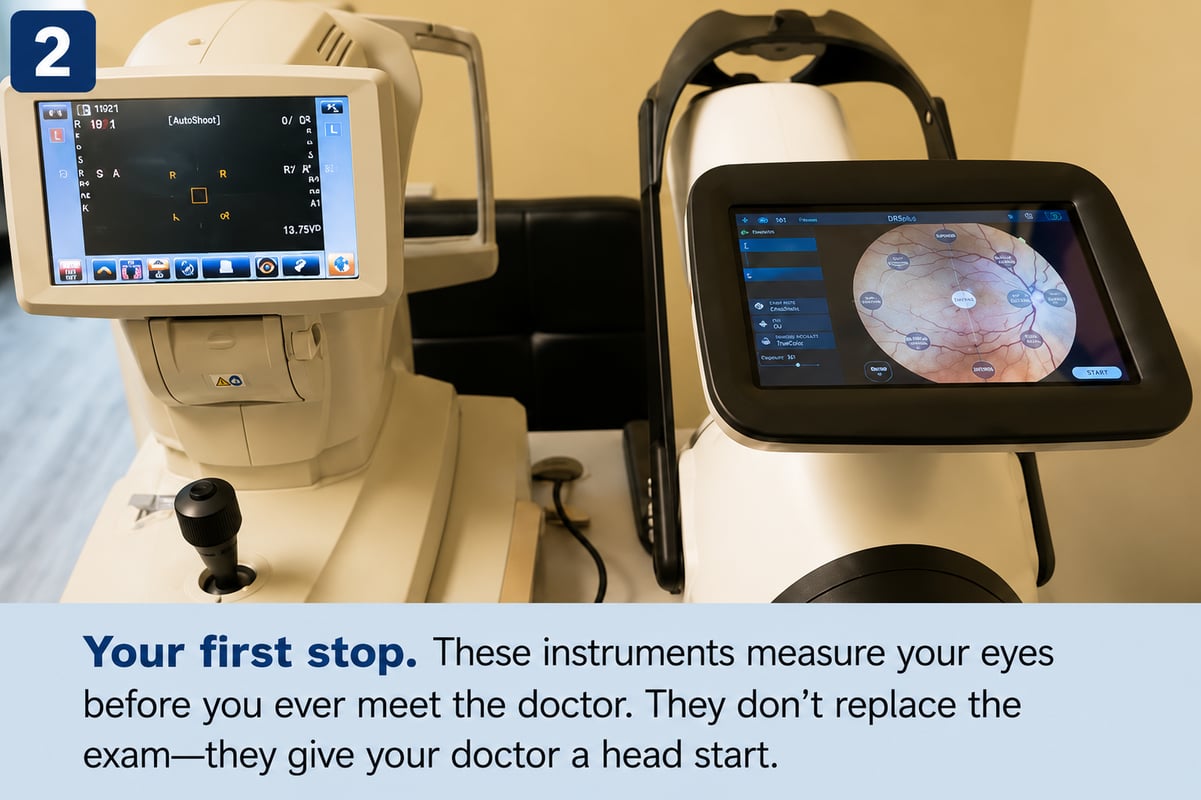
The first stop is our pretesting area. Four measurements, a few minutes, all of them done sitting down with your chin on a rest.
The first machine takes a guess at your prescription. You look at a small picture that drifts in and out of focus while it reads the shape of your eye. Nobody prescribes from that number. It’s a rough draft. What makes it your actual prescription is the conversation we have later, when you tell us which lens looks clearer. Machines are fast. They’re not the final word, and they shouldn’t be.
Then we check the pressure inside each eye, with a device called an iCare tonometer. It touches the front of your eye with a probe so light that most people don’t feel it, and it needs no numbing drops at all. We check it because rising pressure is one of the earliest signs of glaucoma, and glaucoma is quiet. It takes peripheral vision first, so gradually that people don’t notice until a good deal is gone. A number today, compared against a number next year, is how we catch it while it’s still easy to manage. If a reading looks high, or your two eyes differ, we just take it again. One odd number means very little, and we’d rather be sure than alarm you.
Next we read your current glasses. Thirty seconds on a machine tells us the prescription in the pair you walked in wearing. It’s how we can tell whether your eyes have changed much, or whether you’ve simply been overdue for lenses that were never quite right.
Last, we photograph the back of each eye. One bright flash per eye, and we have an image of your retina, your optic nerve, and the blood vessels running across it.
This is the step people are most relieved by: we don’t dilate. No drops, no stinging, no going back to work unable to read your screen, no driving home squinting at three in the afternoon. The camera gives your doctor the detailed look they need without any of that.
Those vessels are worth photographing for their own sake. The back of your eye is the one place in your whole body where blood vessels can be seen directly, without a single incision. Diabetes and high blood pressure leave their marks there, sometimes before anything else shows. Plenty of people have learned something important about their general health from an eye photograph.
In the exam room
Now we slow down. These are the hands-on checks, and I’ll be doing them with you one at a time.
Photographs of the front of your eye. A microscope with a narrow band of bright light lets me photograph your eye in layers: lids, lashes, cornea, the lens inside. Seven images per eye. The light is bright, and I’ll tell you before I turn it on. This is where we find dryness, irritation, early cataract, the small things that explain symptoms people have been putting up with for years.
Following a pen. You keep your head still and track it with your eyes while it traces a wide letter H. Six muscles move each eye, and that pattern walks all of them through their range. It’s how we spot the one that isn’t keeping up, often in someone who’s been getting headaches by mid-afternoon and blaming their screen.
Covering one eye, then the other. I’ll use a small paddle while you look straight ahead. Both eyes can be perfectly sharp on their own and still not aim at quite the same point. When that happens, your brain quietly does extra work all day to hold one image together. That effort has a cost, and it usually shows up as tired eyes in the evening.
Circles full of colored dots. You tell me the number you see in each one. Fourteen plates, a minute at most. Most color vision differences are something you’re born with rather than something you catch, but it’s useful to know, and I’ve watched more than one adult find out for the first time in my chair.
For younger patients, a depth-perception check. Ages 8 to 18, special glasses over their own, and they tell me which shape lifts off the page. When two eyes aren’t teaming up well, it shows up in reading and schoolwork long before a child thinks to mention it. They usually enjoy this one.
Lights down, penlight up. I watch how each pupil answers the light, then swing it between them. Thirty seconds, nothing to do but look ahead. Your pupils sit on a direct line to the nerve behind each eye, and they tell us it’s working.
Meeting your doctor
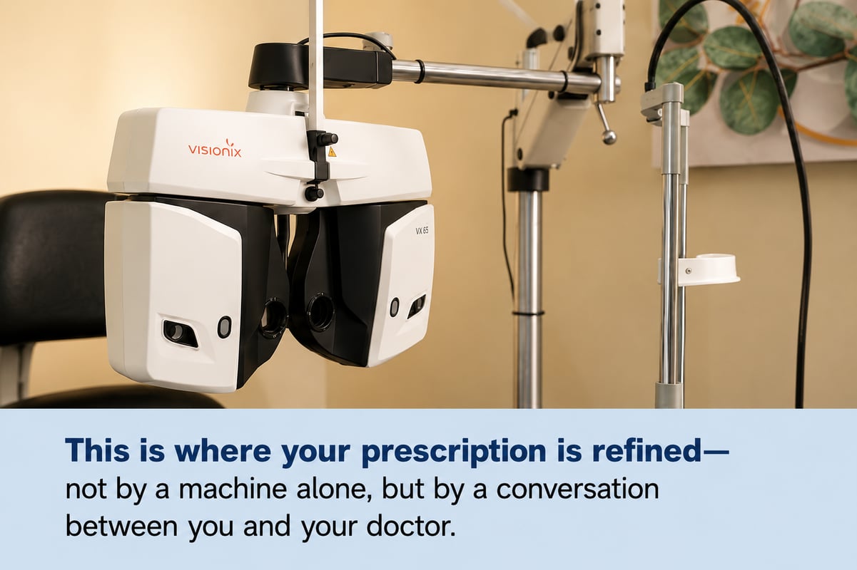
This is where your prescription is made.
Your doctor joins you on a screen in the exam room, and I stay in the room with you the whole time. Every photograph and every measurement we just took is in front of them as you talk.
An eye exam has always been a conversation plus a set of measurements. The measurements are taken here, by me, with my hands. The conversation is with a licensed doctor who is looking at your eyes and listening to your answers in real time. Nothing about your prescription is decided by a machine on its own, and you are never in that room by yourself.
First they’ll check your side vision. One eye covered, and you say when you notice movement at the edges. That’s the vision you use without thinking, the kind that notices a car drifting into your lane.
Then the phoropter swings in front of you. The big lens machine. “Better on one, or two?”
People apologize to me during this part more than any other. They worry they’re getting it wrong, or taking too long, or that the two really do look the same and they’re supposed to see a difference.
“They look the same” is a real answer, and often the most useful one you can give. It tells us we’ve arrived. When the two choices stop being distinguishable, we’re at the edge of what more lens power can do for you, and that’s exactly what we’re listening for. There is no wrong answer. You’re being asked, not tested.
When that’s done, your doctor reviews everything together and finalizes your prescription. If they see anything in your photographs they want you to know about, they’ll tell you then. Ask them whatever you like. That’s the part people wish afterwards they’d used more.
On contact lenses
We do contact lens exams and fittings at our Dallas shop. There’s an extra loop for those: your lenses go back in, we record short videos of how each one sits and moves on your eye, and we measure the vision you’re really getting through them. A lens can be the right power and still be the wrong fit, and that’s what the videos are for.
If contacts are what brought you here, book the contact lens exam rather than the regular one. It runs about forty-five minutes instead of thirty because of the fitting work, and we order your lenses here once your doctor has settled on them.
Then you’re done
The whole thing takes about thirty minutes.
You’ll leave with your prescription in your hand. It’s yours. Fill it with us, fill it anywhere, that has always been your choice and I’ll never make you feel odd for asking. Most people do wander over to the frame wall while they’re already here, and if you’d rather take the paper and think about it, that’s fine too.
Nothing here will hurt
Nothing touches your eye except that feather-light pressure probe. There are no dilation drops. And there’s no such thing as a question that’s too small. If you don’t know why I’m doing something, ask me, and I’ll tell you exactly what I told you here.
You’ve been carrying your eyes around your whole life. Thirty minutes to understand them is not much.
If you’d like to see every step laid out with pictures, we’ve put together a full walkthrough of what to expect at your exam. And when you’re ready, you can book a time that suits you, or just walk in. I’ll be here.
About the Author
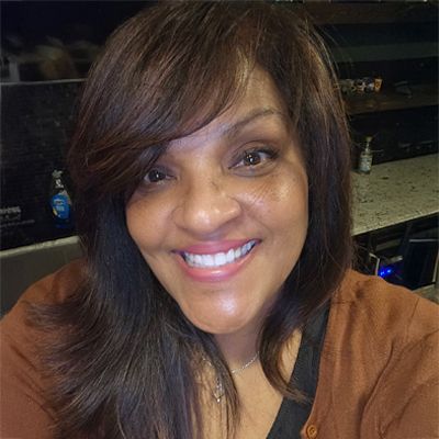
Miriam is a Licensed Optician with more than 30 years of experience helping customers with their vision.